AutoDock Vina
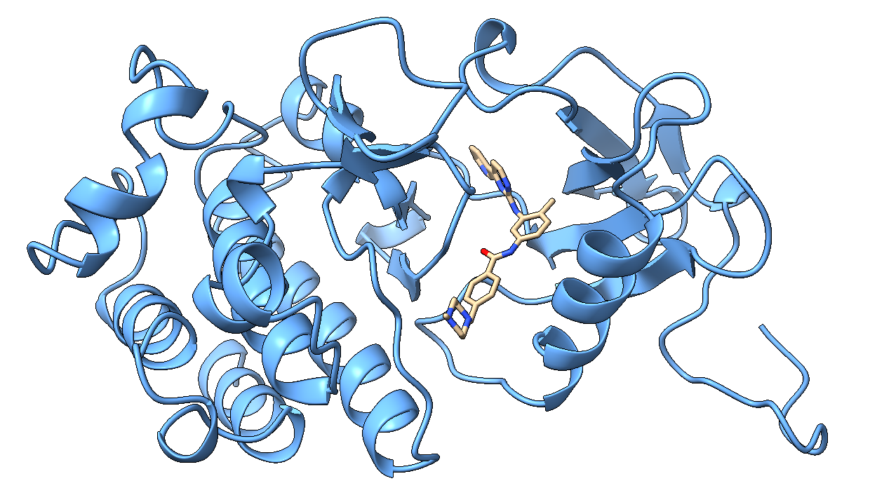
AutoDock Vina is a widely used molecular docking program designed to model the interactions between small molecules (ligands) and macromolecules (receptors). The example presented in this documentation demonstrates the docking of imatinib (Gleevec) into the c-Abl tyrosine kinase protein, an important drug target in the treatment of chronic myeloid leukemia.
Required Software
AutoDock Vina 1.2.7
Meeko
Python 3
AutoGrid4
ChimeraX
Workflow Overview
Receptor preparation
Ligand preparation
Affinity map generation (optional)
Running the docking calculation
Receptor Preparation
The selected structure can be downloaded from
https://www.rcsb.org/3d-view/1IEP.
However, the downloaded structure contains two chains and two ligand molecules, among other components, which makes it unsuitable as a starting structure for our purposes. We require a receptor structure containing hydrogen atoms.
To prepare the input .pdb file, we use ChimeraX. Open the downloaded
1IEP.pdb structure. The receptor can
then be prepared using the following commands in the ChimeraX command line.
First, delete chain B:
delete #1/B
Next, remove everything from chain A that is not part of the protein:
delete ~protein
Then add hydrogen atoms to the resulting receptor:
addh
The resulting structure is now suitable for docking.
The following command can be used to save the prepared receptor (alternatively, the graphical interface can be used):
save 1iep_receptorH.pdb
The file
1iep_receptorH.pdb
will serve as the receptor input structure.
With this file prepared, the receptor can be converted into the format required for docking using the following command:
mk_prepare_receptor.py \
-i 1iep_receptorH.pdb \
-o 1iep_receptor \
-p -v \
--box_size 20 20 20 \
--box_center 15.190 53.903 16.917
This command generates the following files:
1iep_receptor.box.pdb: a file that can be visualized in ChimeraX to display the docking search space1iep_receptor.box.txt: a file containing the search space parameters that can be used as part of the docking configuration
Ligand Preparation
The ligand input file in .sdf format can be downloaded directly from the
Small Molecules section of the
https://www.rcsb.org/structure/1IEP
entry. There, the ligand is listed under the name STI, and the corresponding structure for each chain can be downloaded directly in the desired format.
Hydrogen atoms must be added to the available
1iep_G_STI.sdf ligand before
docking. This can be accomplished in ChimeraX using the same approach described
previously:
addh
The resulting structure is then saved as
1iep_ligand.mol2.
The ligand preparation step can then be performed using the following command:
mk_prepare_ligand.py -i 1iep_ligand.mol2 -o 1iep_ligand.pdbqt
This generates the file:
The official documentation does not recommend the use of the PDB format for small molecules because bond connectivity information may be lost. Instead, SDF or MOL2 formats are preferred.
Generating AutoGrid4 Maps (Optional)
If the AutoDock4 scoring function is to be used, affinity maps must first be generated.
Prepare the receptor together with the GPF file:
mk_prepare_receptor.py -i 1iep_receptorH.pdb -o 1iep_receptor -p -v -g \
--box_size 20 20 20 --box_center 15.190 53.903 16.917
This generates the file
which serves as the input file for AutoGrid4.
autogrid4 -p 1iep_receptor.gpf -l 1iep_receptor.glg
After the calculation, the following files will be created:
1iep_receptor.maps.fld: the main grid file containing references to all affinity maps and a description of the grid geometry1iep_receptor.A.map: affinity map for aromatic/carbon-like atoms1iep_receptor.C.map: interaction-energy map for aliphatic carbon atoms1iep_receptor.N.map: affinity map for neutral nitrogen atoms1iep_receptor.NA.map: affinity map for nitrogen atoms acting as hydrogen-bond acceptors1iep_receptor.OA.map: affinity map for oxygen atoms acting as hydrogen-bond acceptors1iep_receptor.HD.map: affinity map generated for hydrogen-bond donor atoms1iep_receptor.d.map: desolvation map1iep_receptor.e.map: electrostatic map
Docking with the AutoDock Vina Scoring Function
By default, AutoDock Vina uses its own scoring function (Vina scoring function). In this case, precomputed affinity maps are not required; only the receptor PDBQT file and the definition of the search space (grid box) are needed.
vina --receptor 1iep_receptor.pdbqt --ligand 1iep_ligand.pdbqt \
   --config 1iep_receptor.box.txt \
   --exhaustiveness=32 --out 1iep_ligand_vina_out.pdbqt
Important parameters:
--receptor: receptor PDBQT file--ligand: ligand PDBQT file--center_x/y/z: center coordinates of the search space--size_x/y/z: dimensions of the search space in Å--exhaustiveness: thoroughness of the search--out: output file containing the docking results
Larger values of exhaustiveness result in a more extensive search but also
increase the computation time. According to the documentation, imatinib is a
particularly challenging molecule to dock, therefore it is often beneficial to
use a higher value than the default setting of 8.
At the end of the run, the following output is obtained:
mode |   affinity | dist from best mode
     | (kcal/mol) | rmsd l.b.| rmsd u.b.
-----+------------+----------+----------
    1       -13.24          0          0
    2       -11.28      2.996      12.41
    3       -11.14      3.799      12.25
    4        -10.7      1.106      1.622
    5       -10.61      2.572      12.62
    6       -10.27      1.741      13.59
    7       -9.613      3.042      12.57
    8       -9.454      2.363       12.8
    9       -8.981      3.877      12.66
The affinity is reported in kcal/mol. More negative values indicate stronger predicted binding.
Note
The rmsd l.b. and rmsd u.b. values reported by AutoDock Vina do not
represent the RMSD relative to the crystal structure. Instead, they describe
the deviation of each docked pose from the highest-ranked pose (the pose
listed first in the results). Comparison with the experimental crystal
structure requires a separate RMSD calculation.
Docking with the AutoDock4 Scoring Function
One of the new features introduced in AutoDock Vina 1.2.x is the ability to use the AutoDock4 scoring function. In this case, the AutoGrid4 affinity maps generated previously are required.
vina --ligand 1iep_ligand.pdbqt --maps 1iep_receptor --scoring ad4 \
     --exhaustiveness 32 --out 1iep_ligand_ad4_out.pdbqt
Important parameters:
--ligand: ligand PDBQT file--maps: prefix of the maps generated by AutoGrid4--scoring ad4: use the AutoDock4 scoring function--exhaustiveness: search thoroughness--out: output file
When --maps 1iep_receptor is specified, the program automatically loads the
corresponding .maps.fld and .map files.
At the end of the calculation, the following result is reported:
mode |   affinity | dist from best mode
     | (kcal/mol) | rmsd l.b.| rmsd u.b.
-----+------------+----------+----------
    1       -15.21          0          0
    2       -15.07     0.8504      1.045
    3       -13.71      1.291      1.808
    4       -12.72      1.374       2.01
    5       -12.65      3.986      6.062
    6       -11.75      1.629      2.555
    7       -11.25      4.993      11.35
    8       -10.87      3.587      11.47
    9       -10.86       3.55      5.707
Comparison of the Vina and AutoDock4 Force Fields
Vina Scoring Function
Advantages:
simple to use
no need to run AutoGrid4
faster preparation workflow
often a suitable choice for virtual screening
Disadvantages:
may be less accurate for certain ligand-receptor systems
not fully compatible with classical AutoDock4 protocols
AutoDock4 Scoring Function
Advantages:
uses more detailed atom-type-specific energy maps
many older publications and validated protocols are based on it
may provide better reproduction of experimental binding poses for some systems
Disadvantages:
requires additional preparation steps
AutoGrid4 calculations are mandatory
involves managing a larger number of files
It is often worthwhile to dock the same ligand using both scoring functions and compare:
the predicted binding energies,
the resulting binding poses,
key molecular interactions,
RMSD values relative to the crystal structure.
Example:
Method |
Affinity (kcal/mol) |
|---|---|
Vina |
-13.24 |
AD4 |
-15.21 |
The energies should be compared with caution because the two scoring functions are based on different mathematical models. Comparison of the resulting binding poses and key interactions is generally more informative.
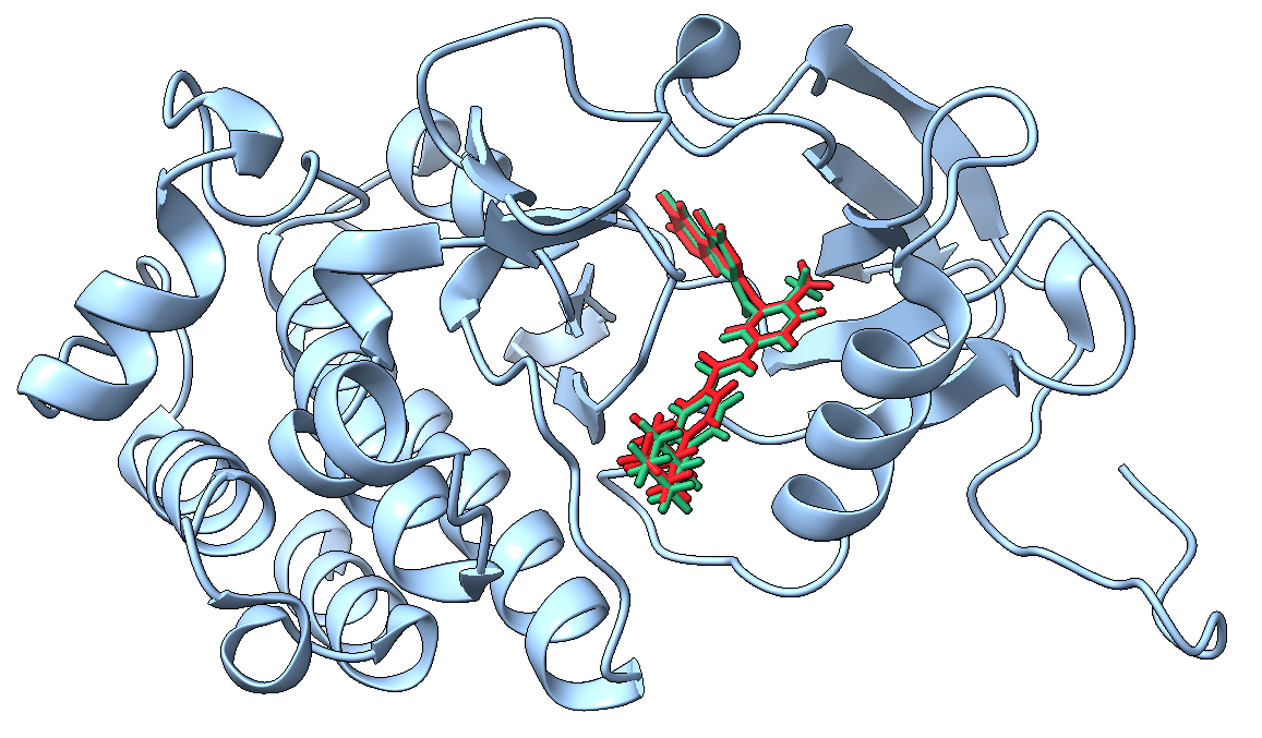
The highest-affinity docking pose obtained using the AutoDock4 force field is shown in red, while the crystal structure is shown in green.
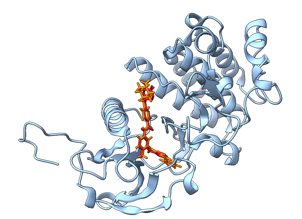
The highest-affinity docking pose obtained using the AutoDock4 force field is shown in red, while the highest-affinity pose obtained using the Vina scoring function is shown in orange.
Generally accepted RMSD values relative to the crystal structure:
RMSD (Å) |
Evaluation |
|---|---|
<2 |
Excellent reproduction |
2-3 |
Acceptable |
>3 |
Significant deviation |
Note
The RMSD between the crystal structure and the docked ligand can only be considered reliable if the atom mapping between the two structures is correct.
Source of the example: https://autodock-vina.readthedocs.io/en/latest/docking_basic.html